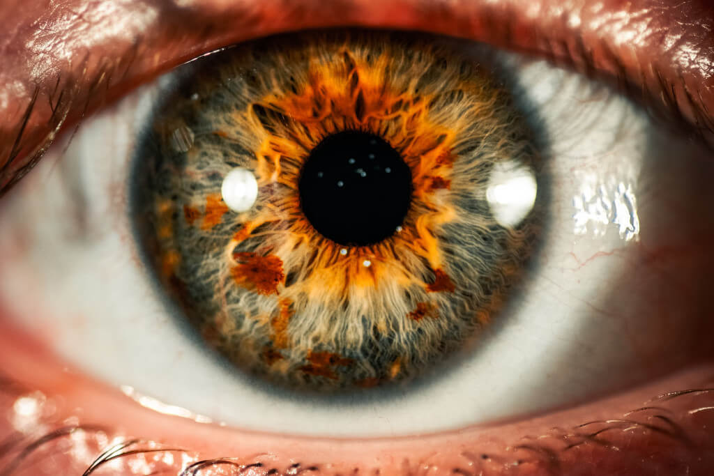
The “Front Window” Of the Eye – The Cornea
The cornea, also known as the straightforward “front window” of the eye, and the natural lens in humans form a complex ocular lens system that serves the same purpose as the lens in a camera. They start concentrating an image on the retina, which is situated in the back of the eye, by bundling parallel light rays that enter the eye (and hence further apart). This image causes the retina to generate an electrical signal, which is then transmitted to the brain via the optic nerve, where it is interpreted and converted into the images we see. The brain creates the images that we see.
The cornea and lens collaborate to determine the focal power of the eye. The cornea accounts for approximately two-thirds of total power, while the lens accounts for approximately one-third of total power. As a result, as you may have realized, the cornea plays an extremely important role in ensuring that a person has a healthy vision. Any imperfection in the cornea, as well as any distortion caused by the cornea, hurts vision.
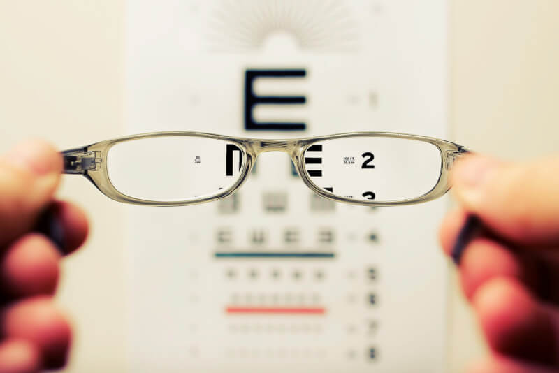
An otherwise healthy eye’s cornea can be reshaped using laser corrective eyewear equipment. This enables the eye to perform its focusing function in an accurate and distortion-free manner.
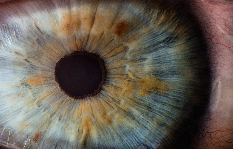
Its Natural Lens
The cornea serves as the “front window” of the eye, allowing light to pass through it and into the retina. It can precisely focus light onto the retina of the eye, which is a very potent ability. It is transparent, sphere-shaped, and made up of five distinct layers.
The epithelium, the most superficial layer, has a high capacity for renewal and recovers quickly from shallow wounds. On the cornea’s periphery, a ring of specialized cells known as limbal stem cells can be seen. These are the cells that are in charge of the corneal epithelium’s regeneration and renewal.
These cells undergo continuous mitosis, also known as cell proliferation, and migrate across the cornea in the direction of both the cornea’s center to replace cells lost as a result of the eyelids’ blinking activity. These cells also form a barrier along the cornea’s border, preventing conjunctival epithelial advancement onto the cornea. This barrier is located at the cornea’s limbus.
The Second Layer
This layer of tissue is the Bowman’s layer, which is followed on the inside by the stroma, Descemet’s membrane, and endothelium. The epithelium is the final layer. The corneal stroma is made up of collagen fibers that are arranged in a geometric pattern, among other things. These fibers form a lattice, and the chemical bonds they share allow them to maintain their highly ordered configuration.
The 3 components in the middle provide stiffness and are designed to allow light to pass through. The cornea’s clarity is due, in part, to the fact that the endothelium’s internal layer is composed of a single layer of cells with the highly specialized feature of keeping the cornea dehydrated. This is just one of the many remarkable properties of the cornea.
The Refractive System
Because it both transmits and bends light, the cornea is an important optical component (like a lens). It is the most prominent piece of the eye’s refractive system, accounting for more than two-thirds of the overall power of the eye to bend light. This directs light into the retina at the back of the eye, resulting in a picture of the original item being created, albeit upside down and much smaller.
For several reasons, the cornea needs to stand out among anatomical structures. In addition to being transparent and the most dehydrated tissue in the human body, it is also the tissue with the most powerful sensory nerve supply. As a result, the brain can tell whether the surface of the cornea has been damaged by dryness, injury, or disease. When the tear film is disrupted or evaporates, these sensory nerves send a signal to the brain, causing the central nervous system to cause tear secretion and the blinking reflex. This maintains the optical quality of the cornea’s external surface, which is required for clear focusing.
Normal Sight
The regular human eye receives light from the left side. The cornea, in conjunction with the natural lens of the eye, works in perfect harmony to properly focus images on the retina, which is located in the back of the eye. These images are crystal clear, sharp, and in focus.
Myopia
Items far away and intermediate in distance appear foggy and out of focus, whereas objects close-up and personal appear sharp and in focus. These people must wear corrective lenses, either spectacles or contact lenses, to see clearly. A near-sighted person’s eye is often longer than a normal person’s eye, and the cornea of their eye may also be steeper. As a result, light is concentrated directly in front of the retina as it passes through the cornea and lens. As a result, photos in the distance will become blurrier. Nearsightedness can be corrected at almost any level by using one of the many refractive surgical treatments that are now available.
Hyperopia

Farsighted people frequently experience difficulty reading up close before the age of 40. A farsighted eye has a flatter cornea than a normal cornea and is slightly shorter than a normal eye. As a result, light from distant objects would then focus behind the retina unless the natural lens can compensate adequately. Nearby objects require a significantly greater capacity for attention to be seen clearly, and as a result, they blur faster. As a result, items close to the lens and intermediate in distance appear blurry and out of focus, whereas objects further away are in focus but not necessarily as sharp as they should be, as demonstrated by the example below. Farsightedness can be corrected surgically using a variety of techniques, including LASIK, refractive lens exchange, and intraocular contact lenses.
Astigmatism
Astigmatism is characterized by an uneven light focus, which occurs when the cornea or natural lens steepens asymmetrically. As a result, the images focus on various locations in front of and/or behind the retina. Individuals with uncorrected astigmatism have blurry vision at all distances, as well as the image of numerous ghost images. Astigmatism is quite common and can coexist with a variety of other refractive errors. To achieve the best results, the treatment should address both the primary and secondary issues, which may not be immediately apparent. This should be accomplished during a single therapy session. Astigmatism can be treated with glasses, contact lenses, corneal relaxing incisions, laser vision adjustment, and customized surgically implanted intraocular lenses.